For Pet Parents
To All Pet Parents
Dr. Josh does all communication through your family veterinarian and does not speak with pet parents directly. This is because your family veterinarian has all of the pieces of the puzzle and your pet’s history and they should always remain in primary control of your pet’s health. This avoids having “too many cooks in the kitchen.”
From Dr. Josh
Dear Pet Parent,
Your family veterinarian has recommended that your loved one have an ultrasound examination. I am an Emergency and Critical Care Specialist, meaning that I have received extra training and education in many aspects of medicine focusing on emergency and critical care, which includes special training with diagnostic ultrasounds. My role in your pet’s health is to help provide additional information and diagnostic support to your veterinarian.

Frequently Asked Questions (FAQs)
What is ultrasound?
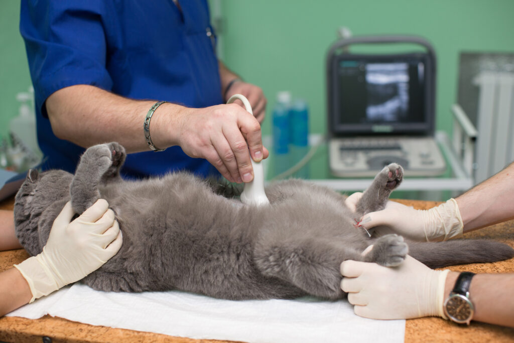
Ultrasound uses harmless sound waves to look “inside” of soft tissues and get a picture of the shape and a comparison of the normal texture or color of various parts of the body. A small probe is placed on your pet’s skin with some gel and the sound waves are transmitted through the skin, with the machine interpreting the echo of that sound wave into a picture. Ultrasound examination is completely harmless and painless for your pet.


What can you expect?
Your pet will need to have the hair shaved away, because air trapped between hairs interferes with the ultrasound signal. Your pet will usually need to be dropped off with your veterinarian for a few hours, because I am travelling to many hospitals in a given day. I cannot always pinpoint the exact time I will arrive to your veterinarian and other pets may also need to have ultrasound examinations, too. Once the examination is completed, I review the results and my opinion verbally with your veterinarian and leave them a report for your pet’s medical record; your veterinarian will then consider this information along with the rest of their knowledge and expertise about your loved one. Your veterinarian will discuss with you the newinformation they have obtained and what the best next steps are for your pet.
How should we prepare ahead of time?
Unless your pet has a medical reason they cannot tolerate fasting (due to things like diabetes or young puppies/kittens), you should try and withhold food after 10pm the night before; food and gas in the stomach blocks some of the areas we want to examine. Water is okay to have available unless otherwise instructed. If you have ANY questions or concerns about this, please ask your veterinarian.
For dogs, please walk them first thing in the morning to reduce the amount of stool that gets in the way. Try to NOT walk your pet within 1 hour of the examination, as we want some urine in the bladder so that we can see this organ better, and have enough there if we need to obtain a sample. Once the study is done, your veterinarian will let you know when your pet can be picked up to go home.

What else should I know?
The ultrasound examination itself generally takes 15 to 30 minutes, depending on their size, shape, and if there are fine details that need to be examined closely. Your pet needs to stay relatively still through the examination and not be panting or breathing too heavily resulting in motion, and therefore MAY need to have some sedation. Most pets do not need sedation and we only use sedative medications in order to keep your pet safe, if they have an overly excited personality and cannot lay still enough to obtain quality information, or to help minimize their stress during the examination, especially if they have a nervous personality. It is important to know that we avoid sedation unless your veterinarian and Dr. Josh agree that it is in your pet ‘s best interests of safety and minimal stress.
Sometimes, we find things on ultrasound that need more information to try and reach a more definitive diagnosis. The most common procedure used during ultrasound is called a fine needle aspirate; this uses a very fine needle to take a few cells from the area of interest and place them on to glass slides for a pathologist to evaluate the type of cells. Aspirates do not hurt more than a minor bug bite or bee sting and most patients barely notice them happening. There is always a risk of bleeding any time a needle is used, however this risk is very low and if bleeding occurs, usually clots on its own. You should be aware that most times when aspirates are performed, we can obtain a more substantial diagnosis; sometimes the results are inconclusive or non-diagnostic because it is only a small sample of a few cells and some pathologic cells do not sample very easily (meaning they hold tight and stay in place). This is a difference from a biopsy, where a chunk of tissue is taken. The reason we use aspirates before taking biopsy samples is that they are generally safer (less risk of bleeding) and are much less invasive.
